
An ultrasound image simply is not a good tool for estimating foetal size, and that applies to both underweight and those who are large for their gestational age. Instead, they often find their predictions to be off by multiple pounds. Foetal Sizeįinally, ultrasounds don’t do not allow doctors to predict a baby’s size with accuracy. Or perhaps the ultrasound takes place a bit too early and not all of their parts have developed, which can lead to an inaccurate determination of their gender. Your baby’s positioning at the time of the gender ultrasound can affect gender predictions. Still, you’ve likely heard stories about how these predictions have been incorrect, and parents only found out when the baby was born. Most of the time, the ultrasound technician can tell your baby’s gender at your second ultrasound, the one at 18 to 21 weeks.

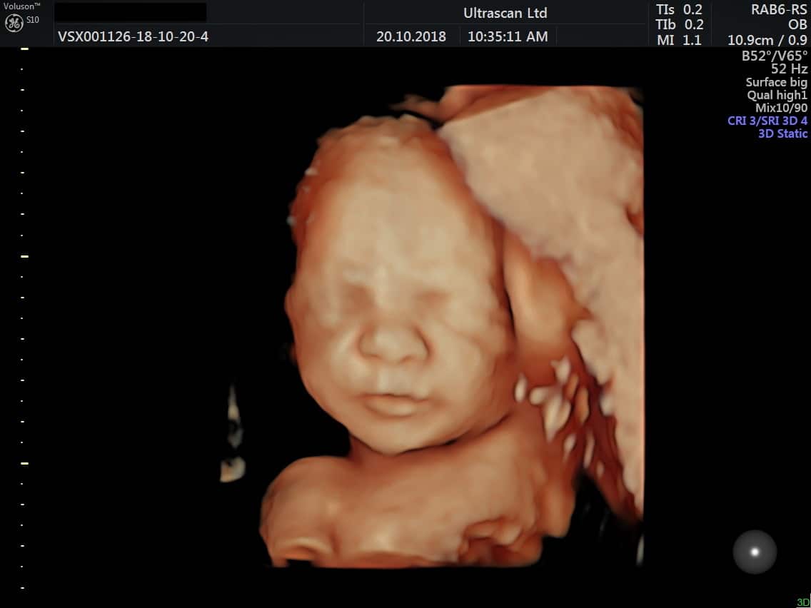
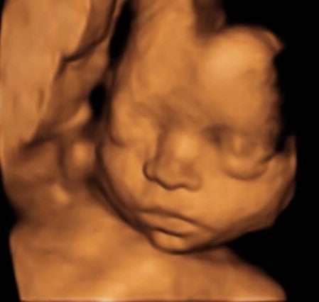
Perhaps you want to stock up on adorable fashions for your little boy or girl. You might want to know your baby’s gender so that you can plan your nursery based on it. Most women will deliver on a day that was not the due date she expected. Surprisingly, only four per cent of expectant mums will go into labour naturally and give birth on that specified day. Regardless of how clear the imaging is, most ultrasounds have a 1.2-week margin of error, meaning your provided due date can be slightly off. But that only applies in the first and second trimesters-due dates shouldn’t change based on an ultrasound after that. Due Date Predictionįor one thing, an ultrasound can help determine a more exact due date for you than tracking your last menstrual period. Results tend to vary most in the following areas. It all depends on the person performing your scan, the machinery they use, the quality of the image, and more. In general, there’s a margin of error when it comes to ultrasound scans. Sonographers can use the images to confirm the baby’s gender and if you’re having multiples, too-the same as they would with a 2D image. As with a normal ultrasound, a 3D scan will incorporate heartbeat and positioning checks. Regardless of what type of scan you choose, your and your baby’s health is of the utmost importance. It’s the same as the 3D scan, except you’ll get a video of the little one growing inside of your womb, in addition to the images of what your baby looks like. These 3D scans result in printed photos that you can take home and share. Nowadays, though, you can have a more accurate scan that provides you with a three-dimensional image of your baby. But that image will be two-dimensional, showing you a basic picture of your little one. A traditional ultrasound provides you a picture into your womb-and of your baby. Prenatal genetic diagnostic tests.Before we dive in, let’s go over the basics of a three-dimensional scan. Amniocentesis.Īmerican College of Obstetricians and Gynecologists. doi:10.1002/j.Īmerican College of Obstetricians and Gynecologists. Australian Journal of Ultrasound in Medicine. Accuracy of sonographic fetal gender determination: Predictions made by sonographers during routine obstetric ultrasound scans. Guidelines for diagnostic imaging during pregnancy and lactation. Ultrasound exams.Īmerican College of Obstetricians and Gynecologists. doi:10.5402/2012/524537Īmerican College of Obstetricians and Gynecologists. Why do parents prefer to know the fetal sex as part of invasive prenatal testing?.


 0 kommentar(er)
